-
E-mail
2820057736@qq.com
-
Phone
13310077902
-
Address
No. 18 Hangdu Road, Yangpu District, Shanghai
Product Categories
Shanghai Zhuocai Biotechnology Co., Ltd
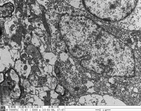
Scanning electron microscopy and transmission electron microscopy
NegotiableUpdate on 01/16
- Model
- Nature of the Manufacturer
- Producers
- Product Category
- Place of Origin
Overview
Complete set of transmission electron microscopy, steps 1 of transmission electron microscopy preparation: Sample fixation: Determine the sampling site of fresh tissue, minimize mechanical damage such as pulling, bruising, and compression, and the tissue volume generally does not exceed 1mm × 1mm × 1mm. Quickly add the electron microscopy fixation solution and fix it at 4 ℃ for 2-4 hours. Centrifuge the cells to the bottom of the tube to see mung bean sized cell clusters. Discard the culture medium and add the electron microscopy fixation solution and fix it at 4 ℃ for 2-4 hours. Rinse three times with 0.1M phosphate buffer PBS (PH7.4) for 15 minutes each time.
Product Details
Complete set of transmission electron microscopy
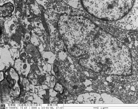
Complete set of transmission electron microscopy
Step 1 of Transmission Electron Microscopy Preparation: Sampling and Fixation: Determine the sampling site for fresh tissue and minimize mechanical damage such as pulling, bruising, and compression. The tissue volume should generally not exceed 1mm × 1mm × 1mm. Quickly add the tissue to the electron microscopy fixative and fix it at 4 ℃ for 2-4 hours. Centrifuge the cells to the bottom of the tube where green bean sized cell clusters can be seen. Discard the culture medium and add the electron microscopy fixative and fix it at 4 ℃ for 2-4 hours. Rinse three times with 0.1M phosphate buffer PBS (PH7.4) for 15 minutes each time. 2. Post fixation: Fix 1% osmium acid in 0.1M phosphate buffer PBS (pH 7.4) at room temperature (20 ℃) for 2 hours. Rinse 3 times with 0.1M phosphate buffer PBS (pH 7.4) for 15 minutes each time. 3. Dehydration: The tissue is dehydrated by ascending with 50% -70% -80% -90% -95% -100% -100% alcohol in sequence, for 15 minutes each time. 4. Penetration: A mixture of acetone: 812 embedding agent=1:1 was infiltrated overnight, while pure 812 embedding agent was infiltrated overnight. 5. Embedding: 60 ℃ polymerization for 48 hours. 6. Slicing: Slicing machine slices ultra-thin sections of 60-80nm. 7. Staining: Uranium lead double staining (2% uranyl acetate saturated aqueous solution, lead citrate, each staining for 15 minutes), slices dried overnight at room temperature. 8. Observe and collect images for analysis under a transmission electron microscope.
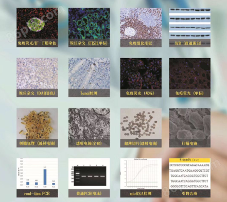
Shanghai Zhuocai Biotechnology Co., Ltd. can undertake various pathological experiments. The samples not only include basic animal tissue slices, but also special samples such as plant leaves, animal cells, bone slices, brain slices, and skin. There are many types of staining and a wide range of staining methods Paraffin staining and ice cutting staining can be performed, and we can also undertake overall experimental projects, cell experimental projects, including sample modeling, cell culture, as well as subsequent experiments on cell proliferation, cell adhesion, cell scratching, etc